US Army X-Ray Field Unit Miscellaneous Medical Equipment

X-Ray Field Unit, Machine, X-Ray, Complete (Item # 96085) ready for shipment at the Picker X-Ray Corporation Factory, Cleveland, Ohio.
Introduction:
Roentgen rays had already been discovered long before the outbreak of WW2. Wilhelm Conrad Röntgen, a German physicist whose discovery would eventually earn him a Nobel Prize, first produced and detected the rays in 1895 whilst working at the University of Würzburg. At first, the findings were called x-rays; but scientists later referred to them as roentgen rays. Soon after their discovery, it was realised that these rays possessed the amazing power of penetrating solids and otherwise opaque media. This power of x-rays distinguishes them from practically all other types of radiation. Professor Röntgen passed the rays through wood, cloth and living tissues during his experiments in November 1895. Later, lead, iron and other heavy materials were shown to be transparent to them in varying degrees. Röntgen demonstrated their property of darkening photographic plates, and that they caused many substances to emit light (i.e. fluorescence). Photographic and fluorescent actions, together with their great penetrating power formed the basis for early applications of the roentgen-ray techniques.
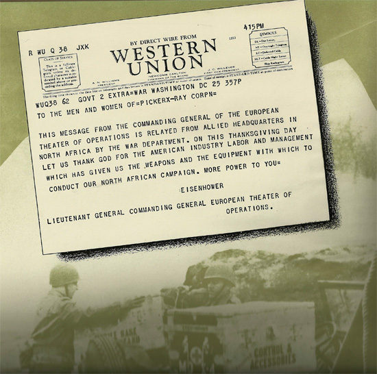
Western Union telegram prepared by Lieutenant General Eisenhower to the men and women of Picker X-Ray Corporation, thanking the company for its labor and management for the war effort.
During the interbellum period, the US Army investigated ways to utilize the new technologies that roentgen rays had presented. The Picker X-Ray Corporation had already been investigating potential applications of the roentgen rays in the medical profession, and the company was approached by the US Army in the late 1930s to produce a special x-ray machine that could be used in the field or in fixed hospitals with very little adjustment.
The Picker X-Ray Corporation of New York was one of the pioneers in the development and manufacture of X-Ray equipment for the U.S. Army. It was awarded the Army-Navy “E” (E for excellence) Flag on 28 August 1942, and received its first star 10 July 1943 and a second star 26 February 1944.
Dr. Henry E. Waite founded the original “Waite and Bartlett Company”, which manufactured medical batteries, static machines, and electrical equipment serving the medical profession. The electro-medical experience of the company would prove extremely valuable in the development of apparatus to put the newly-discovered x-ray apparatus to practical use.
The Waite Company equipped America’s first Hospital Ship, the USS Solace in 1914 with x-ray equipment, and in 1916 the Company working closely together with members of the US Army Medical Division developed and manufactured the original US Army Mobile Bedside X-Ray Unit (first practical use of the hot cathode type x-ray tube).

Another telegram addressed to the men and women of the Picker X-Ray Corporation, this time relaying praise from mobile hospitals in Italy for the Field X-Ray Units the company had provided.
In 1930, the Picker X-Ray Corporation acquired the assets of the Waite and Bartlett Company, and the new group’s achievements greatly influenced the further development of modern x-ray apparatus. Picker-Waite engineers invented, developed, and produced the first completely shock-proof x-ray apparatus as well as the first shock-proof vertical fluoroscope. After an expansion program which took place in 1937, the Company initiated development of a new x-ray unit suited to modern military issues. As it became evident that gasoline-electric generators for proper operation of field equipment were not available, the Picker engineering staff conceived and developed such items for large-scale production during war. Early in the stages of development, transportation became a chief concern, and as result of further study, a strong, compact, and versatile x-ray unit was developed for all emergency situations, yet light enough for convenient transportation by air.
In World War Two, more than 85% of the Picker Corporation production was devoted to field x-ray equipment for the United States Armed Forces, and beside field units, many larger base hospital units were installed in both Army and Navy Hospitals throughout the Zone of Interior.
Picker X-Ray Corporation was the sole manufacturer of x-ray units for the United States Army during WW2. Smaller, less complex items were also produced by other medical laboratory and manufacturing companies, including D. W. Onan & Sons, Inc. (Minnesota) and Westinghouse Electric Company (Pennsylvania). This Article looks at the numerous pieces of field x-ray equipment that were produced exclusively for the US Army during WW2. Each item is presented with its nomenclature, Medical Department Item Number, illustrations and a brief description of the piece.

An X-Ray Technician performs foreign body localization using the X-Ray Field Unit. Notice that he wears a lead apron and a pair of lead-impregnated gloves.
X-Ray Equipment:
Chest, Film, X-Ray (Item # 96025):

X-Ray Field Unit Film Chest (Item # 96025).
This is simply a standard Medical Department Field Chest containing an inner lining of sheet lead. The sheet lead is of such thickness (approximately 1 1/2mm) that, with the opacity of the Chest itself, there is provided protection against x-rays produced by all Army equipment. This special Chest was designed for use particularly at the Evacuation Hospital and for occasional times when films would be needed for use in a truck at the Mobile Surgical Hospital. This particular Chest is not generally used for the shipment of cassettes, cardboard holders or film, since the additional weight of lead lining is unnecessary. As a result, standard Medical Department Field Chests can be used for this purpose.
Dryer and Loading Bin Combination (Item # 96055):

X-Ray Field Unit, Dryer and Loading Bin Combination (Item # 96055).
This unit is intended for General Hospitals, Station Hospitals and Evacuation Hospitals. Occasionally it may be needed for use in a truck. It is portable, being composed of two compartments. The upper compartment is completely lead-lined to afford protection against x-radiation. It consists of two sections: a film storage section occupying the right half, and a loaded cassette section occupying the left half. Racks have been provided in each of these two sections to accommodate the three standard dimensions of x-ray films as used by the Medical Department: 8” x 10”, 10” x 12” and 14” x 17”.
The lower compartment of the unit contains a drawer type of drying rack, a heater, fan and a floor loading shelf. The drying rack has accommodations for 18 films as suspended on standard film hangers. After mounting the upper compartment (the film and cassette loading bins) onto the lower compartment, there is provided a loading bench. The fixation of these two compartments is accomplished by means of a spring-type lock that is found on the inside of the base compartment and beneath its top.
X-Ray Field Unit, Generator (Item # 96060):

X-Ray Field Unit Generator (Item # 96060), weighing less than 400 pounds and capable of supplying the necessary electric energy for the US Army shock-proof X-Ray Field Unit.
This generator is a specially-designed gasoline electrical generator. It is intended particularly for use where community electrical supply is not available and for emergency needs at other hospital installations. Its functioning capacity is such as to permit the delivery of 2,500 watts of energy. The Generator is designed in such a manner that it can be stored inside a modified Medical Department Field Chest.
Grid, Portable (Item # 96070):
This item is a wafer type of grid included with the later models of the Field Table Unit (Item # 96145) and the Fluoroscopic Foreign Body Localization outfit (Item # 96215), or it may be requisitioned separately for general use. The lead strips of this grid are unusually thin. There are 55 strips per inch width of the grid itself – thus even though it is a stationary type, grid marks are inconspicuous. In the case of the first several hundred that were purchased by the Medical Department, the spacing of the lead strips was such as to provide a true centering (grid radius) at 36 inches; later versions were provided for true centering at 30 inches.
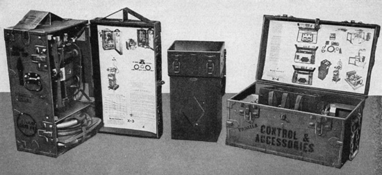
X-Ray Machine Unit (Item # 96085):

X-Ray Field Unit, Machine, X-Ray, Complete (Item # 96085).
This item is intended for all types of hospital installations. It is packed in three Field Chests. Two of these Chests are modified Medical Department Field Chests, the other is of special construction for accommodation of the high tension and filament transformers. The x-ray tube in its housing is packed by setting it in a nest supported by eight springs which are attached to the inside corners of a compartment contained in one of the Field Chests. In addition, this particular Chest accommodates a drum for packing the two shockproof cables, as well as smaller items such as the filters (to be used in roentgenotherapy) and the several aperture diaphragms. This Chest has a gable type of superstructure which has been added in order to emphasize that the chest should be placed on end during transit. This is important in order to provide the greatest protection to the x-ray tube.
The second standard Medical Department Field Chest is modified for accommodation of the Control Unit, the interconnecting cables, the timer and the foot switch. A lead apron, a pair of lead-impregnated gloves and a pair of goggles can also be accommodated in this Chest.
Complete X-Ray Field Unit packed in Medical Department Field Chests ready for transportation.
In conjunction with the X-Ray Table Unit (Item# 96145) or the Mobile X-Ray Chassis (Item # 96090), this machine unit is adaptable nine ways: horizontal roentgenoscopy, foreign body localization by means of a rapid roentgenoscopic method, sitting roentgenoscopy, standing roentgenoscopy (to the extent of accommodating routing chest studies and also gastrointestinal studies), horizontal roentgenography (using focal film distances up to 45 inches), 6-foot vertical chest studies, 6-foot horizontal chest studies, ordinary bedside work on wards and superficial roentgenotherapy.
The item is broken down into a number of separate components, each with a specially designated Medical Chest (MD X-#) as follows:
Item # 96086 –X-Ray Field Unit Transformer Unit, Chest – MD X-2
Item # 96087 –X-Ray Field Unit Tube Unit, Chest – MD X-1
Item # 96088 – X-Ray Field Unit Control Unit, Chest – MD X-4
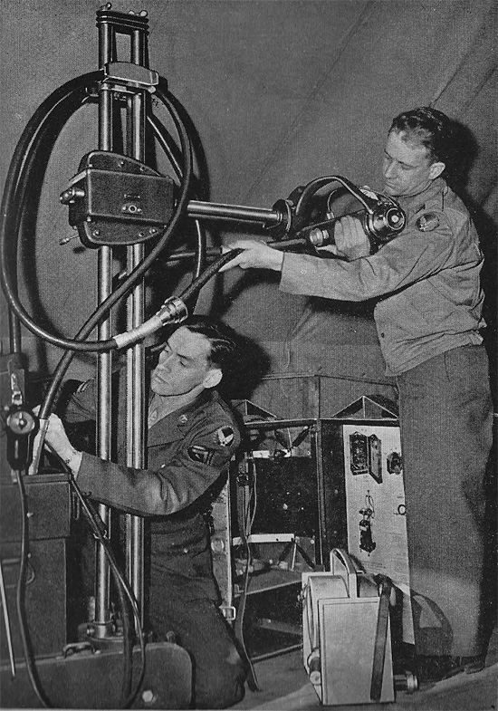
Tec 5 Ned Wolfe and Pvt. William Sullivan, detailed to the Medical Detachment at Mitchel Field demonstrate the assembly of the X-Ray Field Unit. Complete assembly of the unit was possible in eight minutes.
Additional replacement items are available for the unit: X-Ray Field Unit, Shockproof Cable (Item # 96205), X-Ray Field Unit, Tube, X-Ray (Item # 96209), and X-Ray Field Unit, Kit Spare Parts and Repair, X-Ray Machine (Item # 96245). It should be noted that from March 1944 onwards, the Control Unit was produced in two variants, one for the standard 110 volt power supply, and another, which was capable of handling 110-220 volts. These were designated Item # 9608808 and Item # 9608810 respectively.
Mobile Chassis Unit (Item # 96090):

Item # 96085 assembled with Item # 96090 showing the adaptation as a mobile X-Ray Machine Unit. The unit represents the assembled contents of 3 Medical Chests, plus the transformer unit (Item # 96086), which only takes five minutes to set up.
This special chassis has been designed to convert the X-Ray Machine Unit into a mobile unit for use in the wards and at the bedside. It should be of service in the hospitalization section of the Mobile Surgical Hospital, in the Evacuation Hospital, the General Hospital and the Station Hospital. An additional model is provided with wheels of greater diameter and with means of pulling the X-Ray Machine Unit when mounted on it, as well as pushing it. These additional features have been incorporated to facilitate maneuvering the equipment over rough terrain (such as is typically found in the field). This second model is designated Item # 96090-10.

Chassis for Field Use (Item # 96090-10).
Processing Unit (Item # 96115):
This item has been developed for use in any type of hospital installation. It is designed as two sections; a Master Chemical Section (Item # 96115) and an Auxiliary Wash Tank (Item # 96117)When using the master chemical section alone, 30 to 50 large films can be accommodated within an 8-hour period. With the addition of the Auxiliary Wash Tank, this can be increased to 200 or more films.
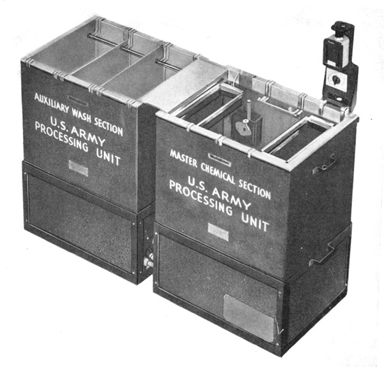
X-Ray Field Unit Processing Unit, for Darkroom (Item # 96115) with Auxiliary Wash Tank (Item # 96117).
The Master Chemical Section consists of a tank compartment and a pedestal or base compartment. The tank compartment has a volume capacity of approximately 50 gallons. The pedestal or base compartment contains a water-circulator unit, a refrigerator unit, a heating unit, a mixing chamber and a thermostatic regulator. The unit is assembled by simply setting the tank compartment upon the pedestal compartment and connecting three water couplings which can be accomplished without the need of tools. The electrical supply required is 115 volts, 50-90 cycle, single-phase. This can be provided by the Gasoline Electrical Generator unit (Item # 99600).
The Auxiliary Wash Tank consists of two compartments; a tank compartment which is identical in dimensions with those of the Master Chemical Tank, and a pedestal compartment. The tank compartment contains two baffles to provide for a cascade type of washing. A pre-cooling system is packed in the pedestal compartment. When in use, the pre-cooling coils are suspended in the tank of the Master Chemical Section.
Both sections of the Processing Unit are composed of two compartments. This provision is for the sake of portability, making the unit practical both for permanent installations and for use in the field. In case of necessity it can be carried in parts to a location where activities may require it. The heating and cooling units as well as the pre-cooler are self-contained and all connections can be made by hand, without the use of any special tools.
X-Ray Table Unit (Item # 96145):

X-Ray Field Unit Table Unit (Item # 96145) assembled.
This item is a portable roentgenoscopic and roentgenographic table unit. It is considered practical for usage in any type of hospital installation. Essentially, this unit is composed of two fabricated steel end-pieces, three rails (each composed of three sections), a horizontal carriage and a C-shaped supporting arm for accommodation of the x-ray tube unit and also of the fluoroscopic screen.

Item # 96145 packed in Chests ready for transportation.
The C-shaped arm may be adjusted in several ways:
- The x-ray tube and fluoroscopic screen may be positioned so as to provide for horizontal roentgenoscopy, with an allowance of vertical shifting through a range of 16 inches.
- They may be positioned so as to provide for vertical roentgenoscopy, the patient being in the sitting position.
- They may be positioned to one end of the table chassis so as to provide for standing roentgenoscopy.
- The tube housing itself may be rotated to a position so as to provide for chest studies with focal film distances as great as or greater than 6 feet.
- In the position for conventional horizontal roentgenography, the tube housing may be raised or lowered so as to provide focal film distances varying from 29 inches to as much as 45 inches, the patient lying on a litter at the table-top level.
- The x-ray tube may be rotated in the horizontal plane so as to provide for chest studies of focal film distances as great as 6 feet, the patient lying upon a litter placed on its feet.
Darkroom Tent (Item # 96175):

X-Ray Field Unit, Darkroom Tent (Item # 96175).
The Tent has been designed for usage particularly at mobile hospitals where it is expected that it will be needed both with the mobile operating section and with the hospitalization section. Its design is such that it can be erected indoors in any room (8 by 10 feet in area and 8 feet in height) as well as within a tent or dugout or by itself, in the open. It is therefore quickly adaptable for use in a temporary building or any building taken over for service as an Evacuation Hospital. It may even serve a purpose at a General Hospital, though at this installation it is more likely that substantial darkroom construction will be had.

By adjusting the inner curtain of the Tent, it was possible to convert the space from a darkroom to a film processing laboratory easily.
Its design includes a two-way adaptation for roentgenoscopy and / or for film processing. It provides for a quick changeover where lightproof conditions are essential. When erected outdoors, additional support may be obtained by weighting apron with turf, rocks or other heavy ballast. Further support may be provided by utilizing auxiliary guy ropes. The guy ropes are not included with the item, but eyelets are provided. The Tent is otherwise supported by means of a sectional frame, which is contained within a specially-designed Chest.
X-Ray Machine and Table Unit (Item # 96215):

X-Ray Machine and Table Unit (Item # 96215) assembled for horizontal roentgenography.
This unit has been designed primarily for horizontal roentgenoscopy, horizontal roentgenography and roentgenoscopic localization of foreign bodies. The equipment is so-designed that it can be packed into three lightweight chests for ease of transportation across difficult terrain. The packing arrangement is ingeniously provided for in order to accommodate all the component parts of the equipment. To simplify assembly, printed identifications and color codes are provided on all parts. The Unit consists of the table component, shockproof head and supporting hanger, control, and automatic protective and auxiliary devices. A spare parts compartment for tools and miscellaneous parts is provided in the Control Unit. A spare tube is also provided as a component of the unit. For roentgenoscopy and foreign body localization, a fluoroscopic screen, depth scale marker and localization dial together with necessary accessories are included.
The generating circuit of the unit may be connected to 100-130 or 200-240 volts, 50-60 cycle alternating current. The Control Unit is compact and it contains all the features incorporated into Item # 96085 with modifications and refinements. The shockproof head includes the tube, high tension transformer, and filament transformer immersed in oil. This arrangement obviates the necessity for shockproof cables. Incorporated within the head is the oil impeller and air circulating motors. In addition, it permits the placement of the shockproof components into one insulated head. The x-ray tube of this unit is accommodated in a separate compartment within the shockproof head so that easy exchange of it can be accomplished without the usual difficulties concerned with removing and replacing transformer oil.
The table portion of the unit is assembled with the use of special tools provided and in accordance with instructions printed on the inside of the lids of the Chests. The table is also provided with a cassette lifting device for carrying out roentgenography.
X-Ray Equipment Literature:
Additional data, descriptions, and use of X-Ray Field Units and Equipment can be found in TM 8-275 “Military Roentgenology”, dated January 26, 1943, and TM 8-632 “X-Ray Field Unit, Machine, Chassis & Table, Items 9608508, 9608510, 9609005, 9609010, and 9614500” dated February 1945.